Table of Contents
- Non-invasive Imaging Biomarker of EGFRvIII in Glioblastoma Patients
- Prediction of Overall Survival in Glioblastoma Patients
- Probability Maps of Potential Recurrence of Glioblastoma Tumors
- Prediction of Progression-Free Survival (PFS) and Recurrence Pattern (RP) in Glioblastoma
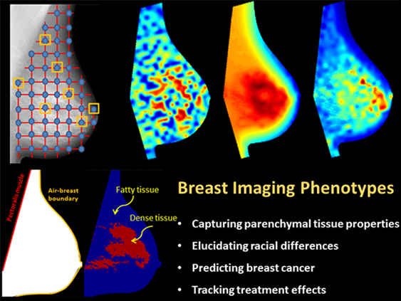
- Imaging Biomarkers Related to Cancer Risk and Development of Breast Cancer
This section presents examples of applications using CaPTk.
Please make sure that whenever you use and/or refer to CaPTk in your research, you should always cite the following papers:
- C.Davatzikos, S.Rathore, S.Bakas, S.Pati, M.Bergman, R.Kalarot, P.Sridharan, A.Gastounioti, N.Jahani, E.Cohen, H.Akbari, B.Tunc, J.Doshi, D.Parker, M.Hsieh, A.Sotiras, H.Li, Y.Ou, R.K.Doot, M.Bilello, Y.Fan, R.T.Shinohara, P.Yushkevich, R.Verma, D.Kontos, "Cancer imaging phenomics toolkit: quantitative imaging analytics for precision diagnostics and predictive modeling of clinical outcome", J Med Imaging, 5(1):011018, 2018, DOI:10.1117/1.JMI.5.1.011018
- S.Pati, A.Singh, S.Rathore, A.Gastounioti, M.Bergman, P.Ngo, S.M.Ha, D.Bounias, J.Minock, G.Murphy, H.Li, A.Bhattarai, A.Wolf, P.Sridaran, R.Kalarot, H.Akbari, A.Sotiras, S.P.Thakur, R.Verma, R.T.Shinohara, P.Yushkevich, Y.Fan, D.Kontos, C.Davatzikos, S.Bakas, "The Cancer Imaging Phenomics Toolkit (CaPTk): Technical Overview", Springer - BrainLes 2019 - LNCS, Vol.11993, 380-394, 2020, DOI:10.1007/978-3-030-46643-5_38
In addition, if the journal/conference where you submit your paper does not restrict you from citing abstracts you might also cite the following:
- RRID: SCR_017323
- S.Rathore, S.Bakas, S.Pati, H.Akbari, R.Kalarot, P.Sridharan, M.Rozycki, M.Bergman, B.Tunc, R.Verma, M.Bilello, C.Davatzikos. "Brain Cancer Imaging Phenomics Toolkit (brain-CaPTk): An Interactive Platform for Quantitative Analysis of Glioblastoma", BrainLes 2017. LNCS Springer, 10670:133-145, 2017, DOI:10.1007/978-3-319-75238-9_12
- S.Pati, S.Bakas, A.Sotiras, R.Kalarot, P.Sridharan, M.Bergman, S.Rathore, H.Akbari, P.Yushkevich, T.Shinohara, Y.Fan, D.Kontos, R.Verma, C.Davatzikos. "Cancer Imaging Phenomics Toolkit (CaPTk): A Radio(geno)mics Software Platform Leveraging Quantitative Imaging Analytics for Computational Oncology", 103rd Scientific Assembly and Annual Meeting of the Radiological Society of North America (RSNA), Nov.26-Dec.1, 2017, Chicago IL.
- S.Pati, S.Rathore, R.Kalarot, P.Sridharan, M.Bergman, T.Shinohara, P.Yushkevich, Y.Fan, R.Verma, D.Kontos, C.Davatzikos. "Cancer and Phenomics Toolkit (CaPTk): A Software Suite for Computational Oncology and Radiomics", 102nd Scientific Assembly and Annual Meeting of the Radiological Society of North America (RSNA), Nov.27-Dec.2, 2016, Chicago IL. archive.rsna.org/2016/16014589.html
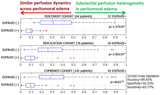
Non-invasive Imaging Biomarker of EGFRvIII in Glioblastoma Patients
References:
- S.Bakas, H.Akbari, J.Pisapia, M.Martinez-Lage, M.Rozycki, S.Rathore, N.Dahmane, D.M.O'Rourke, C.Davatzikos, "In vivo detection of EGFRvIII in glioblastoma via perfusion magnetic resonance imaging signature consistent with deep peritumoral infiltration: the phi-index", Clin Cancer Res. 23(16):4724-4734, 2017, DOI:10.1158/1078-0432.CCR-16-1871
- S.Bakas, H.Akbari, J.Pisapia, M.Rozycki, D.M.O'Rourke, C.Davatzikos. "Identification of Imaging Signatures of the Epidermal Growth Factor Receptor Variant III (EGFRvIII) in Glioblastoma", Neuro Oncol. 17(Suppl 5):v154, 2015, DOI:10.1093/neuonc/nov225.05
- S.Bakas, Z.A.Binder, H.Akbari, M.Martinez-Lage, M.Rozycki, J.J.D.Morrissette, N.Dahmane, D.M.O'Rourke, C.Davatzikos, "Highly-expressed wild-type EGFR and EGFRvIII mutant glioblastomas have similar MRI signature, consistent with deep peritumoral infiltration", Neuro Oncol. 18(Suppl 6):vi125-vi126, 2016, DOI:10.1093/neuonc/now212.523
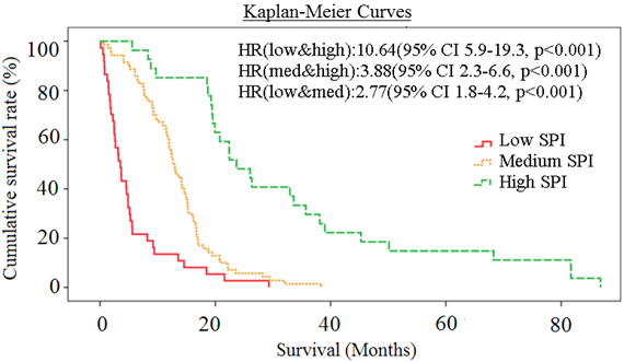
Prediction of Overall Survival in Glioblastoma Patients
Reference:
- L.Macyszyn, H.Akbari, J.M.Pisapia, X.Da, M.Attiah, V.Pigrish, Y.Bi, S.Pal, R.V.Davuluri, L.Roccograndi, N.Dahmane. M.Martinez-Lage, G.Biros, R.L.Wolf, M.Bilello, D.M.O'Rourke, C.Davatzikos. "Imaging patterns predict patient survival and molecular subtype in glioblastoma via machine learning techniques", Neuro Oncol. 18(3):417-25, 2016, DOI:10.1093/neuonc/nov127
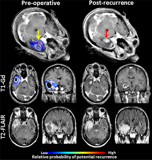
Probability Maps of Potential Recurrence of Glioblastoma Tumors
References:
- H.Akbari, L.Macyszyn, X.Da, M.Bilello, R.L.Wolf, M.Martinez-Lage, G.Biros, M.Alonso-Basanta, D.M.O'Rourke, C.Davatzikos. "Imaging Surrogates of Infiltration Obtained Via Multiparametric Imaging Pattern Analysis Predict Subsequent Location of Recurrence of Glioblastoma", Neurosurgery. 78(4):572-80, 2016, DOI:10.1227/NEU.0000000000001202
- H.Akbari, L.Macyszyn, X.Da, R.L.Wolf, M.Bilello, R.Verma, D.M.O'Rourke, C.Davatzikos, "Pattern analysis of dynamic susceptibility contrast-enhanced MR imaging demonstrates peritumoral tissue heterogeneity", Radiology. 273(2):502-10, 2014, DOI:10.1148/radiol.14132458
- H.Akbari, L.Macyszyn, J.Pisapia, X.Da, M.Attiah, Y.Bi, S.Pal, R.Davuluri, L.Roccograndi, N.Dahmane, R.Wolf, M.Bilello, D.M.O'Rourke, C.Davatzikos, "Survival Prediction in Glioblastoma Patients Using Multi-parametric MRI Biomarkers and Machine Learning Methods", American Society of Neuroradiology, O-525:2042-2044, 2015.
Prediction of Progression-Free Survival (PFS) and Recurrence Pattern (RP) in Glioblastoma
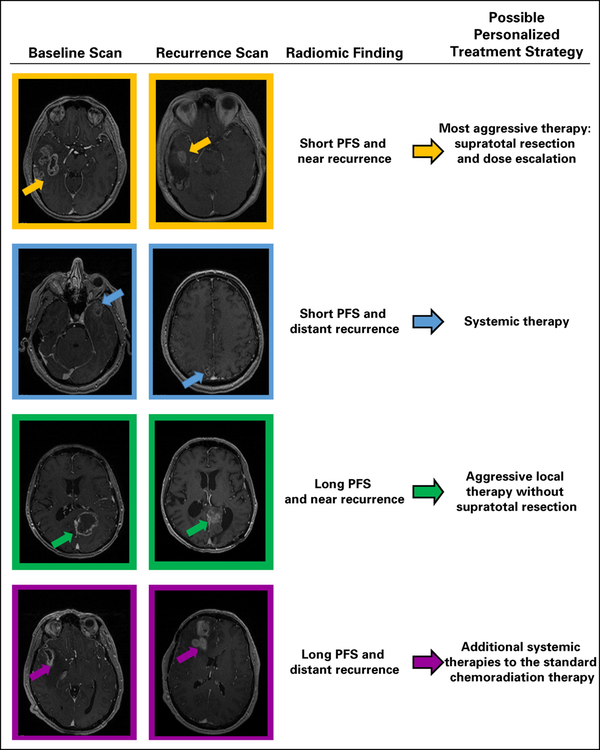
In this study, we investigated the application of in-vivo MP-MRI phenomic signatures leveraging ML and the CaPTk software suite for prediction of PFS and RP in patients with GBM who received standard of care therapy, aiming to offer advanced imaging-based biomarkers for clinical decision making and personalized treatment planning. We showed that predictive radiomic models can be used to help in upfront decision making, on a patient-specific basis. The following image, indicates examples of different schemes of PFS and recurrence pattern with possible personalized treatment strategies: the first and second columns indicate the baseline and recurrence scans for each example, the radiomic finding for each example is displayed in the third column, and the fourth column shows the suggested personalized therapy plan for each example.
References:
- Fathi Kazerooni, Anahita, Hamed Akbari, Gaurav Shukla, Chaitra Badve, Jeffrey D. Rudie, Chiharu Sako, Saima Rathore et al. "Cancer Imaging Phenomics via CaPTk: Multi-Institutional Prediction of Progression-Free Survival and Pattern of Recurrence in Glioblastoma." JCO Clinical Cancer Informatics 4 (2020): 234-244.
- Kazerooni, Anahita Fathi, Saima Rathore, Hamed Akbari, Jeffrey Rudie, Chiharu Sako, Sung Min Ha, Elizabeth Mamourian et al. "QUANTITATIVE ESTIMATION OF PROGRESSION-FREE SURVIVAL BASED ON RADIOMICS ANALYSIS OF PREOPERATIVE MULTI-PARAMETRIC MRI IN PATIENTS WITH GLIOBLASTOMA." Neuro-oncology 21, no. Supplement_6 (2019).
Imaging Biomarkers Related to Cancer Risk and Development of Breast Cancer
References:
- A.Gastounioti, A.Oustimov, B.M.Keller, L.Pantalone, M.K.Hsieh, E.F.Conant, D.Kontos. "Breast parenchymal patterns in processed versus raw digital mammograms: A large population study toward assessing differences in quantitative measures across image representations", Med Phys. 43(11):5862-77, 2016, DOI: 10.1118/1.4963810
- A.M.McCarthy, B.M.Keller, L.M.Pantalone, M.K.Hsieh, M.Synnestvedt, E.F.Conant, K.Armstrong, D.Kontos. "Racial differences in quantitative measures of area and volumetric breast density", J Natl Cancer Inst. 108(10), 2016, DOI:10.1093/jnci/djw104
- E.F.Conant, B.M.Keller, L.Pantalone, A.Gastounioti, E.S.McDonald, D.Kontos. "Agreement between Breast Percentage Density Estimations from Standard-Dose versus Synthetic Digital Mammograms: Results from a Large Screening Cohort Using Automated Measures", Radiology. 283(3):673-80, 2017, DOI:10.1148/radiol.2016161286
- A.D.Williams, A.So, M.Synnestvedt, C.M.Tewksbury, D.Kontos, M.K.Hsieh, L.Pantalone, E.F.Conant, M.Schnall, K.Dumon, N.Williams, J.Tchou. "Mammographic breast density decreases after bariatric surgery" Breast Cancer Res Treat. 165(3):565-572, 2017, DOI:10.1007/s10549-017-4361-y